In a new work published in Cell Reports, researchers used lymph node infections to map Zika virus infections to learn how the virus enters the body through several skin entry points and spreads throughout the body.
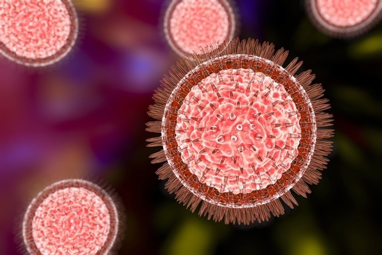
The Flavivirus genus includes the positive-sense, single-stranded RNA virus known as the Zika virus, which is spread by mosquitoes. Recent investigations have revealed that Zika virus infections can cause microcephaly and congenital neurodevelopmental illness, even if the majority of Zika virus infections have been moderate, with some patients demonstrating persistent viremia instances. This has sped up research on the causes of Zika virus infections, possible treatments, and potential vaccinations. Nevertheless, there is presently no approved treatment for or vaccination against the Zika virus.
In the present study, the researchers conducted footpad inoculation of the interferon alpha receptor 1 (Ifnar1–/–) mouse model with the Zika virus. The early stages of the viral delivery kinetics were observed in the popliteal lymph node (PLN), which drains in the hindfoot. The reasoning was that if skin cells were required for viral replication, high viral titers would not be detected in the PLN.
After inoculation with 104 focus-forming units of Zika virus, PLNs were harvested at multiple time points within one hour to determine the viral titers through focus-formation assays. The infectious virus titers from serum samples and PLNs were also quantified every four hours for the first 32 hours post-infection.
Five distinct macrophage populations are found in lymph nodes related to specific immune functions. Macrophages in the nodal sinuses closest to the incoming lymph vessels are called subcapsular sinus macrophages, while those in the nodal sinuses near the exiting lymph vessels are called medullary sinus macrophages.